What Tests Can Be Performed During an ENT Specialist Assessment?
ENT Diagnostic Tests for Ear, Nose and Throat Symptoms
An appointment with an ear, nose and throat specialist is often more detailed than a routine medical consultation. An ENT doctor, also called an otolaryngologist, examines connected structures that influence hearing, balance, breathing, smell, voice, and swallowing.
A symptom in one area may originate somewhere else, so the assessment is designed to build a complete picture rather than focus on a single complaint.
Not every patient needs every test. The specialist chooses investigations according to your symptoms, medical history, and findings during the physical examination. Some checks are completed in the consultation room within minutes. Others require an audiologist, laboratory, imaging department or follow-up appointment.
What Happens Before ENT Testing Begins?
The assessment normally starts with a detailed conversation. You may be asked when your symptoms began, whether they affect one or both sides, how they have changed, and what treatments you have already tried.
The specialist may review ear infections, sinus problems, allergies, previous surgery, head injuries, noise exposure, medication, smoking, reflux, and family history. For dizziness, you may be asked about the length of each episode, triggers, and associated hearing symptoms.
A physical examination usually follows. The doctor may inspect your ears, nose, mouth, throat, neck, and facial movements before deciding whether further tests are necessary.
Otoscopy and Microscopic Ear Examination
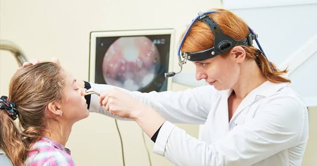
An ENT assessment often begins with a direct look inside the ear. To perform this examination, the doctor uses an otoscope, a compact instrument equipped with illumination and a viewing system that makes the ear canal and eardrum visible.
The examination can help identify several possible reasons for ear discomfort or changes in hearing. These include hardened earwax, irritated canal skin, swelling, abnormal secretions, and visible signs of infection. The doctor can also look for damage to the eardrum, evidence of previous injury, or changes that may suggest fluid has accumulated in the middle ear.
When ordinary viewing does not provide enough detail, additional magnification may be useful. An ENT specialist may examine the ear through a microscope or use a video-based instrument that provides an enlarged view of the structures being assessed.
If wax or discharge prevents adequate examination, the specialist may recommend carefully removing the obstruction. This can sometimes involve microsuction, which uses controlled suction to clear material from the ear canal. Although cleaning helps the clinician inspect the ear more thoroughly, it is considered a treatment or examination-supporting procedure rather than a diagnostic investigation in itself.
Pure-Tone Audiometry for Measuring Hearing
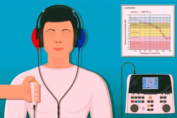
Pure-tone audiometry is a formal hearing test usually conducted in a quiet room or sound-treated booth. You wear headphones and respond whenever you hear tones at different pitches and volume levels.
The test identifies the softest sounds you can hear and produces an audiogram showing hearing thresholds in each ear. It can help determine whether hearing loss is conductive, meaning sound is being blocked in the outer or middle ear, or sensorineural, involving the inner ear or hearing nerve.
Speech Audiometry and Word Recognition Testing
Hearing everyday speech involves more than detecting simple tones. Speech audiometry measures how softly you can recognise spoken words and how accurately you understand them at a comfortable volume.
During word recognition testing, you repeat words presented through headphones. A person may hear that a sound is present yet struggle to distinguish speech, especially in background noise. Comparing speech scores with the pure-tone audiogram provides useful information about hearing function.
Tympanometry and Middle-Ear Pressure Testing
Tympanometry evaluates how the eardrum moves as air pressure changes inside the ear canal. A soft probe is placed at the ear opening, creating a brief sealed sensation. You may hear a tone and feel pressure, but you do not need to respond.
The test can suggest fluid behind the eardrum, reduced eardrum movement, Eustachian tube dysfunction, a perforation, or problems involving the middle-ear bones. It does not directly measure how well you hear; instead, it assesses the mechanical condition of the middle ear.
Acoustic Reflex and Otoacoustic Emission Tests
Acoustic reflex testing measures automatic responses of tiny middle-ear muscles to sound. It can add information about the middle ear, hearing nerve, and pathways involved in sound processing.
Otoacoustic emission testing records faint sounds produced by healthy sensory cells in the inner ear. A small probe delivers tones and measures the response. The patient remains still and does not need to press a button.
Auditory Brainstem Response Testing
An auditory brainstem response test measures electrical activity along the hearing pathway after sounds are played through earphones. Small sensors are attached to the scalp and skin near the ears.
The test can estimate hearing sensitivity when a conventional hearing test is unreliable or impossible. It may also help investigate certain differences between the ears or possible problems affecting the hearing nerve.
Nasal Endoscopy for Blockage and Sinus Symptoms
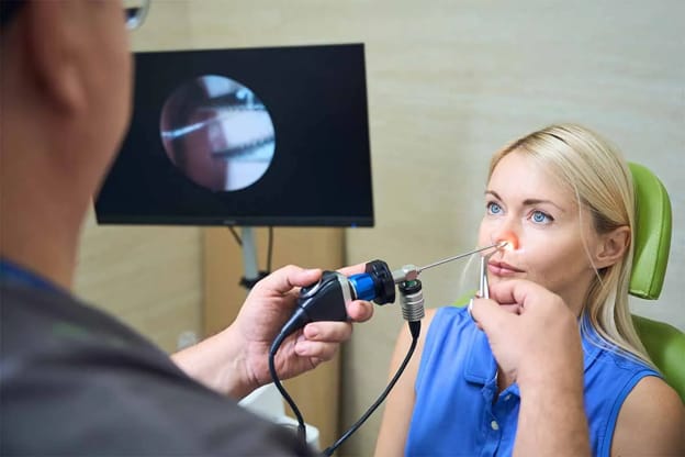
Certain problems affecting the nose and sinuses may develop in areas that are difficult to assess through a routine nasal examination. To investigate these regions more thoroughly, an ENT specialist may perform nasal endoscopy, a procedure that provides a magnified view of the internal nasal structures, sinus drainage pathways, and the space behind the nose, known as the nasopharynx.
During the examination, the specialist carefully guides a slender instrument, known as an endoscope, into the nasal passage. Depending on the patient’s symptoms and the areas requiring assessment, either a flexible or rigid endoscope may be used.
Before the procedure, a nasal decongestant or local anaesthetic spray may be administered to minimise discomfort, reduce tissue swelling, and improve visibility.
By examining the nasal passages directly, the specialist can look for abnormalities that might otherwise remain undetected. These may include persistent inflammation, nasal polyps, excessive mucus, crusting, areas of bleeding, structural narrowing, or unusual tissue growths.
The procedure also allows closer inspection of regions where the sinuses drain into the nose, helping the doctor investigate possible causes of recurring sinus infections, prolonged nasal congestion, unexplained discharge, or difficulty breathing through one nostril.
Although the idea of having an instrument inserted into the nose may seem uncomfortable, nasal endoscopy is generally well tolerated. Patients may notice a temporary feeling of pressure, mild irritation, watery eyes, or an urge to sneeze as the instrument moves through the nasal cavity. The examination typically takes only a few minutes and is usually performed during an outpatient consultation.
If an area of concern is identified, the ENT specialist may recommend additional investigations to determine its nature. In selected cases, a small tissue sample may be collected for laboratory examination through a procedure known as a biopsy.
The findings from nasal endoscopy can help establish a more accurate diagnosis and guide further treatment according to the patient’s condition.
Flexible Laryngoscopy for the Throat and Voice Box
Persistent hoarseness, throat discomfort, swallowing difficulty, chronic cough or a lump sensation may be investigated with flexible laryngoscopy. A slim camera passes through the nose to view the throat, voice box and vocal cords.
The specialist may ask you to breathe, speak, make particular sounds or swallow. This shows how the vocal cords move and can reveal inflammation, weakness, nodules, polyps, reflux-related changes, or another lesion.
A numbing spray may be used. Most people tolerate the examination well, although it can cause brief gagging, coughing, or nasal discomfort. More detailed voice assessment may involve stroboscopy, which uses pulsed light to examine vocal-cord vibration.
Balance and Vestibular Testing
Dizziness does not always originate from an ear problem. Maintaining balance requires coordination between the inner ear, eyes, brain, and nervous system. Disruptions involving these structures, certain medications, or underlying medical conditions can cause spinning sensations, unsteadiness, or difficulty maintaining balance.
During an ENT consultation, the specialist may assess the patient’s symptoms, eye movements, coordination, and balance to identify possible causes. If dizziness occurs when changing head positions, a Dix-Hallpike test may be performed to investigate benign paroxysmal positional vertigo (BPPV), a condition associated with displaced calcium crystals within the inner ear’s balance canals.
The examination involves carefully positioning the patient’s head and body while observing for unusual eye movements, known as nystagmus, and any accompanying spinning sensations. These responses can help identify BPPV affecting the posterior semicircular canal. Additional positional assessments may be necessary if other balance canals are suspected.
For more complex or persistent symptoms, the ENT specialist may recommend videonystagmography (VNG), caloric testing, video head impulse testing (vHIT), or vestibular evoked myogenic potentials (VEMP). Each investigation evaluates different aspects of the balance system, helping the doctor determine the likely source of dizziness and recommend suitable treatment.
Smell and Taste Testing
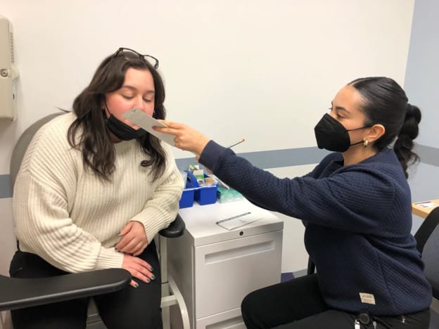
Reduced smell may follow infection, chronic sinus inflammation, nasal polyps, head injury or neurological disease. Standardised smell tests may ask you to identify or compare familiar odours.
Taste problems are less common than perceived loss of taste caused by reduced smell. A specialist may test sweet, salty, sour or bitter sensations when a genuine taste disorder is suspected.
Swallowing Assessments
Swallowing problems may be assessed in the clinic by observing mouth, tongue and throat movements. The specialist may watch you swallow liquids or foods of different textures.
A fibreoptic endoscopic evaluation of swallowing uses a flexible camera passed through the nose while you eat and drink small amounts. It shows whether material collects in the throat or enters the airway.
Another option is a videofluoroscopic swallow study, a moving X-ray performed while you swallow contrast material. This test examines the coordinated movement of food from the mouth through the throat and helps guide dietary or rehabilitation advice.
Allergy Tests, Cultures and Biopsies
Skin-prick or blood tests may be arranged when allergic rhinitis is suspected. Results must be interpreted alongside symptoms because a positive result does not always prove that an allergen is causing the current problem.
A swab or culture may be taken from persistent ear discharge or selected nasal infections, particularly when standard treatment has failed. Laboratory results can identify organisms and guide medication choices.
A biopsy removes a small tissue sample for microscopic examination. It may be recommended for an unexplained ulcer, neck lump, vocal-cord lesion, or nasal growth. Being offered a biopsy does not automatically mean cancer is present; it is a method of obtaining a definite diagnosis.
When Are CT, MRI or Ultrasound Scans Needed?
Imaging is not necessary for every ENT complaint. CT scanning provides detailed views of bone, sinuses, middle-ear spaces and certain neck structures. MRI gives more information about soft tissue, nerves and the brain.
Ultrasound is commonly used to assess thyroid nodules, salivary glands and neck lumps, and it may guide a needle biopsy. The specialist chooses imaging according to the suspected condition and avoids scans that are unlikely to change management.
How Should You Prepare for an ENT Assessment?
Bring a medication list, previous test results, and details of earlier treatment. Note when symptoms occur, what triggers them, and whether they affect one side.
Final Thoughts
An ENT specialist assessment may involve simple inspection, hearing tests, pressure measurements, endoscopy, balance testing, swallowing studies, laboratory analysis, or imaging. The exact combination depends on the problem being investigated.
These tests are not performed merely to collect information. Each one answers a specific clinical question, narrows possible causes, and helps the specialist recommend suitable treatment. Asking what a test is designed to show can make the process clearer and help you take an active role in decisions about your care.